Study sheds light on how picosecond lasers work in pigmentation disorders and skin ageing
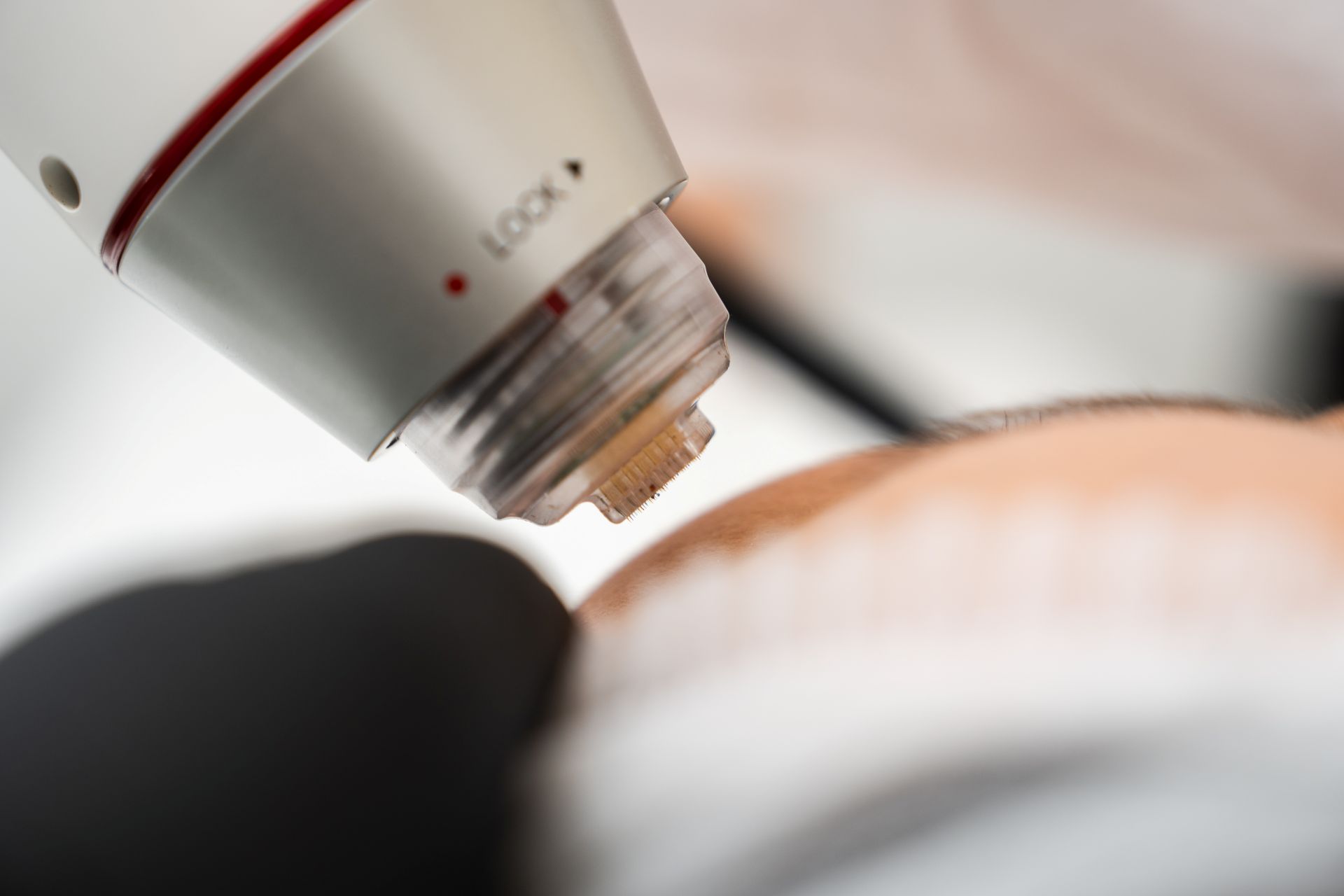
A new preclinical study using a porcine skin model has provided insight into the biological mechanisms underpinning the clinical effects of picosecond lasers, offering data that could support safer parameter selection and more rational use of the technology in dermatology.
The research examined how picosecond lasers affect pigmentation disorders and skin ageing at tissue, cellular and molecular levels. The porcine model was selected because of its close anatomical and physiological similarity to human skin, allowing researchers to explore laser-induced effects in a clinically relevant setting.
According to the study, picosecond lasers reduce pigmentation by fragmenting melanosomes into smaller particles that can be cleared by immune cells, while also suppressing the production of new pigment. At the same time, laser exposure triggered changes associated with skin rejuvenation, including collagen remodelling and increased expression of proteins involved in maintaining the epidermal barrier.
The findings were reported in a basic science article titled Decoding the mechanisms of pigment reduction and skin rejuvenation induced by picosecond laser: insights from a porcine model. The study was published in Lasers in Surgery and Medicine and selected as the journal’s January 2026 editor’s choice.
Xiang Wen, dermatologist and professor at West China Hospital, said the work addresses a gap between  widespread clinical use and limited mechanistic understanding. “Picosecond lasers are widely used in clinical practice for pigmentary disorders and skin rejuvenation, yet their biological mechanisms remain incompletely understood,” Wen said.
She added: “We conducted this study to systematically elucidate how picosecond laser treatment achieves pigment clearance and promotes skin rejuvenation at the tissue, cellular, and molecular levels.”
Analysis of treated skin showed that picosecond laser exposure led to melanosome fragmentation and macrophage-mediated pigment clearance. Researchers also observed a sustained inhibition of melanogenesis, linked to melanocyte loss and reduced expression of tyrosinase, a key enzyme involved in melanin synthesis. Together, these changes help explain the durable pigment-lightening effects seen in clinical practice.
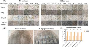
Beyond pigment reduction, the study demonstrated effects relevant to skin ageing. Laser treatment promoted dermal collagen remodelling and upregulation of growth factors associated with tissue repair. In the epidermis, researchers reported increased levels of barrier-related proteins, suggesting an improvement in skin structure and resilience following treatment.
The authors concluded that these combined effects support the dual therapeutic role of picosecond lasers in both pigmentation disorders and skin rejuvenation. By clarifying how different biological pathways are affected, the data may help clinicians optimise treatment parameters to maximise benefit while minimising risk.
While the study was conducted in an animal model, its use of porcine skin provides translational relevance, given its similarity to human skin in thickness, collagen structure and healing response. The researchers noted that a clearer understanding of laser–tissue interactions could support more evidence-based clinical application of picosecond laser technology.